Personalized Treatment Planning for Radiotherapy
Automation software that enables cancer clinics to do more in less time. Transform your department with increased plan quality, safety, and efficiency.
*All website prices include full taxes.

Automation from Start to Finish
Contour
deep-learning contours
Plan
Check
Treat
Personalized Cancer Care
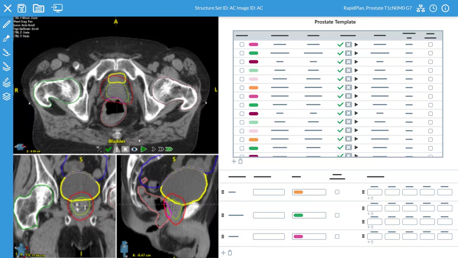
AutoContour comes ready-to-go with 200 CT & MR structure models, from head to femur, ensuring rapid implementation for treatment planning-ready contours in minutes. With no user configuration required, faster contours are just a click away.

This is just a simple text made for this unique and awesome template, you can replace it with any text.

Head & Neck
- Brain
- Brainstem
- Buccal Mucosa
- Cervical Vertebrae (C1-C7)
- Cochlea (L/R)
- Cornea (L/R)
- Ear Internal (L/R)
- Eye (L/R)
- Hippocampus (L/R)
- Lacrimal (L/R)
- Larynx 4
- Lens (L/R)
- Lips
- Macula (L/R)
- Mandible
- Neck Lymph Nodes 20
- Optic Chiasm
- Optic Nerve (L/R)
- Oral Cavity
- Oral Cavity Ext
- Parotid (L/R)
- Pharyngeal Constrictors
- Pituitary
- Retina (L/R)
- Skull
- Submandibular (L/R)
- Temporal Lobe (L/R)
- Thyroid
Thorax
- Aorta (Full/Asc/Desc)
- Axillary Lymph Nodes RTOG 10
- Brachial Plexus (L/R)
- Breast (L/R)
- Bronchus
- Carina
- Chestwall 5
- Esophagus
- Heart
- Humerus (L/R)
- Internal Mammary Lymph Nodes 4
- LAD Artery
- Lung (L/R)
- Nipple (L/R)
- Pericardium
- Pulmonary Artery
- Ribs 3
- Sternum
- Supraclavicular Lymph Nodes 4
- Thoracic Vertebrae (T1-T12)
- Trachea
- Vena Cava Inferior
- Vena Cava Superior
Abdomen
- Aorta (Full/Asc/Desc)
- Body
- Bowel 4
- Duodenum
- Esophagus
- Gallbladder
- Kidney (L/R)
- Kidney Minus Hilum (L/R)
- Liver
- Pancreas
- Spleen
- Spinal Canal
- Spinal Cord
- Stomach
- Thoracic Vertebrae (T01-T12)
- Vena Cava Inferior
- Vertebrae (All)
Pelvis
- Bladder (M/F)
- Bowel 4
- Cauda Equina
- Femur 6
- Genitals (M/F)
- HDR Cylinder
- Iliac Crest (L/R)
- Iliac Marrow (L/R)
- Inguinofemoral Lymph Nodes (L/R)
- Lumbar Vertebrae (L1-L5)
- Paraaortic Lymph Nodes
- Pelvic Bones
- Pelvic Lymph Nodes
- Pelvic Lymph Nodes NRG
- Penile Bulb
- Prostate
- Rectum (M/F)
- Seminal Vesicles
- Sigmoid Colon
- Utero Cervix
MR Models
- Prostate
- Prostate Gland
- Seminal Vesicles

Elevate your diagnostic capabilities with HeadNeckPro, the AI-powered assistant crafted for the early identification and assessment of head and neck cancers. Through sophisticated imaging analysis, it swiftly draws attention to abnormalities, providing a graded suspicion level that guides radiologists in prioritizing cases and optimizing patient outcomes.”
The head and neck squamous cell carcinoma (HNSCC) is the most common cancer in men in India. The current study is a single-center hospital based retrospective study to understand the epidemiology of HNCs in terms of demographic and clinical characteristics at the time of diagnosis and their survival analysis.

MammoX: Tailored for Radiologist Precision and Efficiency. Crafted to seamlessly integrate into the radiologist’s workflow, MammoX delivers intuitive, actionable insights. With Saige Dx at its core, it assigns a distinct Suspicion Level to each finding and the overall case, clearly delineating the likelihood of cancer from Minimal (MIN) to Low (LO), Intermediate (INT), and High (HI), ensuring critical decisions are made with utmost confidence and accuracy
Finding suspicion level
The Finding Suspicion Level represents the strength of suspicion that a given region of interest (outlined by a bounding box) is malignant. Depending on the case, there may be no findings or there may be multiple. Each finding is assigned its own Suspicion Level.
Case suspicion level
The Case Suspicion Level indicates the strength of suspicion that the overall case contains at least one malignant finding. The MammoX algorithm combines information from all processed images and findings into a single Suspicion Level.
This is just a simple text made for this unique and awesome template, you can replace it with any text.
